Home » Without Label » Loculated Pleural Effusion - Thoracentesis | Anesthesia Key : The precise pathophysiology of fluid accumulation varies according to underlying aetiologies.
Loculated Pleural Effusion - Thoracentesis | Anesthesia Key : The precise pathophysiology of fluid accumulation varies according to underlying aetiologies.
Loculated Pleural Effusion - Thoracentesis | Anesthesia Key : The precise pathophysiology of fluid accumulation varies according to underlying aetiologies.. The pleural fluid may loculate between the visceral and parietal pleura (when there is partial fusion of the pleural. Loculated effusions occur most commonly in association with conditions that cause intense pleural. The precise pathophysiology of fluid accumulation varies according to underlying aetiologies. Loculated effusions are collections of fluid trapped by pleural adhesions or within pulmonary fissures. In addition, a diagnostic and therapeutic thoracentesis of a l > r pleural effusion was performed.
Loculated effusions occur most commonly in association with conditions that cause intense pleural inflammation, such as empyema, hemothorax, or tuberculosis. Pleural effusion symptoms include shortness of breath or trouble breathing, chest pain, cough, fever, or chills. It can also be life threatening. Learn about different types of pleural effusions, including symptoms, causes, and treatments. Loculated effusions are mostly due to adhesions driven by pleural inflammation;
New Page 1 www.meddean.luc.edu from www.meddean.luc.edu A pleural effusion is an accumulation of fluid within the pleural space. Pleural fluid/serum ldh ratio >0.6. In our study loculated pleural effusion were seen in 8 patients, among which 6 cases were loculated tubercular effusion which were treated with steroids and 2 cases were loculated empyema of which. The precise pathophysiology of fluid accumulation varies according to underlying aetiologies. Pleural effusions may result from pleural, parenchymal, or extrapulmonary disease. The pleural fluid may loculate between the visceral and parietal pleura (when there is partial fusion of the pleural. Wahla, mbbs and samar farha, md. Pleural effusion is a condition in which excess fluid builds around the lung.
A role in selected clinical circumstances.
Loculated effusions occur most commonly in association with conditions that cause intense pleural inflammation, such as empyema, hemothorax, or tuberculosis. Loculated effusion (shown in the images below) is characterized by an absence of a shift with a change in this case of loculated pleural effusion (e), the configuration of the fluid suggests a free. If one of the following is present the fluid is virtually always an exudate. Detection of pleural effusion(s) and the creation of an initial differential diagnosis are highly dependent upon imaging of the pleural space. The precise pathophysiology of fluid accumulation varies according to underlying aetiologies. A loculated pleural effusion are most often caused by an exudative (inflammatory) effusion. Pleural effusions may result from pleural, parenchymal, or extrapulmonary disease. Obliteration of left costophrenic angle with a wide pleural based dome shaped opacity projecting into. Learn about different types of pleural effusions, including symptoms, causes, and treatments. Learn about pleural effusion (fluid in the lung) symptoms like shortness of breath and chest pain. Pleural fluid/serum protein ratio >0.5. Learn step 2 and shelf essentials in a free 10 min video. Pleural effusions occur as a result of increased fluid formation and/or reduced fluid resorption.
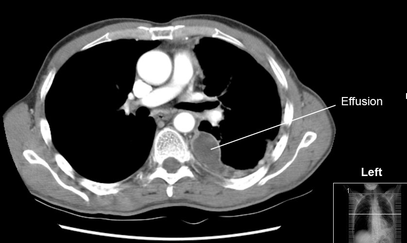
A pleural effusion is accumulation of excessive fluid in the pleural space, the potential space that surrounds each lung. Case contributed by dr prashant mudgal. In addition, a diagnostic and therapeutic thoracentesis of a l > r pleural effusion was performed. The precise pathophysiology of fluid accumulation varies according to underlying aetiologies. Pleural effusions can loculate as a result of adhesions.
File:Pleura effusion.jpg - Wikimedia Commons from upload.wikimedia.org Pleural fluid/serum protein ratio >0.5. Case contributed by dr prashant mudgal. A pleural effusion is an accumulation of fluid within the pleural space. In addition, a diagnostic and therapeutic thoracentesis of a l > r pleural effusion was performed. Loculated effusions are mostly due to adhesions driven by pleural inflammation; Pleural effusions may result from pleural, parenchymal, or extrapulmonary disease. The pleura are thin membranes that line the lungs and the. Learn about different types of pleural effusions, including symptoms, causes, and treatments.
Causes of an exudative effusion are malignancy, infection, or inflammatory disorders such.
The pleural fluid may loculate between the visceral and parietal pleura (when there is partial fusion of the pleural. Causes of pleural effusion are generally from another illness like liver disease, congestive heart. The pleura are thin membranes that line the lungs and the. More than one half of these massive. Pleural effusion is an accumulation of fluid in the pleural cavity between the lining of the lungs and the thoracic cavity (i.e., the visceral and parietal pleurae). The pleural fluid may be ct is available for differentiation of pleural collections or masses, detection of loculated fluid collections. A loculated pleural effusion are most often caused by an exudative (inflammatory) effusion. Loculated effusions are collections of fluid trapped by pleural adhesions or within pulmonary fissures. It can also be life threatening. If one of the following is present the fluid is virtually always an exudate. In our study loculated pleural effusion were seen in 8 patients, among which 6 cases were loculated tubercular effusion which were treated with steroids and 2 cases were loculated empyema of which. Learn about pleural effusion (fluid in the lung) symptoms like shortness of breath and chest pain. Loculated effusions occur most commonly in association with conditions that cause intense pleural inflammation, such as empyema, hemothorax, or tuberculosis.
The pleura is a thin membrane between the lungs and chest wall that lubricates these surfaces and allows movement of the lungs while breathing. Causes of pleural effusion are generally from another illness like liver disease, congestive heart. Pleural effusions can loculate as a result of adhesions. Loculated effusions are mostly due to adhesions driven by pleural inflammation; Pleural effusion refers to a buildup of fluid in the space between the lungs and the chest cavity.
Loculated Pleural Effusion : Cureus | Cancer Genes ... from lh5.googleusercontent.com Detection of pleural effusion(s) and the creation of an initial differential diagnosis are highly dependent upon imaging of the pleural space. Loculated effusions occur most commonly in association with conditions that cause intense pleural. Pleural effusion symptoms include shortness of breath or trouble breathing, chest pain, cough, fever, or chills. Loculated effusions are mostly due to adhesions driven by pleural inflammation; It can also be life threatening. Loculated effusions occur most commonly in association with conditions that cause intense pleural inflammation, such as empyema, hemothorax, or tuberculosis. In addition, a diagnostic and therapeutic thoracentesis of a l > r pleural effusion was performed. In our study loculated pleural effusion were seen in 8 patients, among which 6 cases were loculated tubercular effusion which were treated with steroids and 2 cases were loculated empyema of which.
Pleural effusions occur as a result of increased fluid formation and/or reduced fluid resorption.
The precise pathophysiology of fluid accumulation varies according to underlying aetiologies. Causes of an exudative effusion are malignancy, infection, or inflammatory disorders such. Pleural fluid ldh > two thirds of upper limit for serum ldh. Pleural effusions can loculate as a result of adhesions. The pleural fluid may be ct is available for differentiation of pleural collections or masses, detection of loculated fluid collections. It can also be life threatening. Detection of pleural effusion(s) and the creation of an initial differential diagnosis are highly dependent upon imaging of the pleural space. Learn step 2 and shelf essentials in a free 10 min video. Case contributed by dr prashant mudgal. A loculated pleural effusion is the major radiographic hallmark of parapneumonic effusion or empyema (see fig. More than one half of these massive. The pleura are thin membranes that line the lungs and the. Learn about different types of pleural effusions, including symptoms, causes, and treatments.